Brillouin microscopy of the zebrafish eye
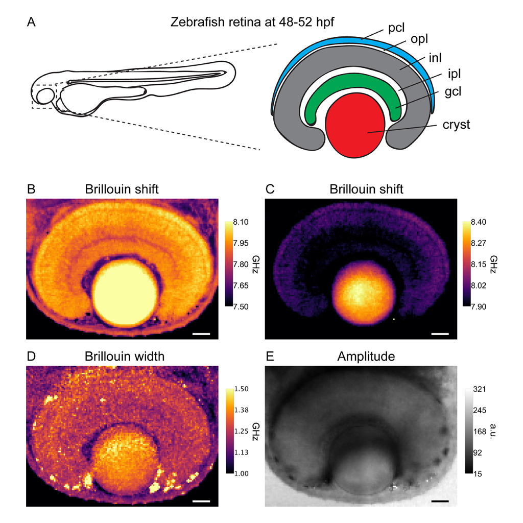
The study explores the mechanical properties of the developing eye in zebrafish larvae using three-dimensional Brillouin Microscopy, an advanced, non-invasive optical technique. This approach allows researchers to measure elasticity and viscosity by analyzing the Brillouin scattering spectra from the eye tissues, including the crystalline lens and various retinal layers. Capturing data from zebrafish embryos 48–52 hours post-fertilization, the research provides a detailed understanding of the eye’s complex longitudinal modulus, linking mechanical properties to developmental biology. This insight is crucial for dissecting the mechanobiological aspects of vertebrate eye development and offers a fundamental starting point for further investigation into how mechanical forces influence organogenesis.
The experimental procedure involved using a confocal Brillouin Microscope equipped with a dual-stage virtually imaged phase array spectrometer, which finely resolves the spectral data necessary for this analysis. Zebrafish larvae were prepared and imaged under carefully controlled conditions to ensure high precision in data collection while minimizing potential damage from the imaging process. The spectral data was meticulously analyzed using custom software, enabling the researchers to extract meaningful mechanical metrics from the high-resolution images. The study’s findings enhance the understanding of the mechanical heterogeneity across different neuronal layers in the vertebrate retina, providing valuable data that could influence future research in tissue engineering and regenerative medicine.
Source and Copyright:
Sánchez-Iranzo, Héctor, et al. “A 3D Brillouin Microscopy dataset of the in-vivo zebrafish eye.” Data in Brief 30 (2020): 105427.
https://doi.org/10.1016/j.dib.2020.105427
https://creativecommons.org/licenses/by/4.0/