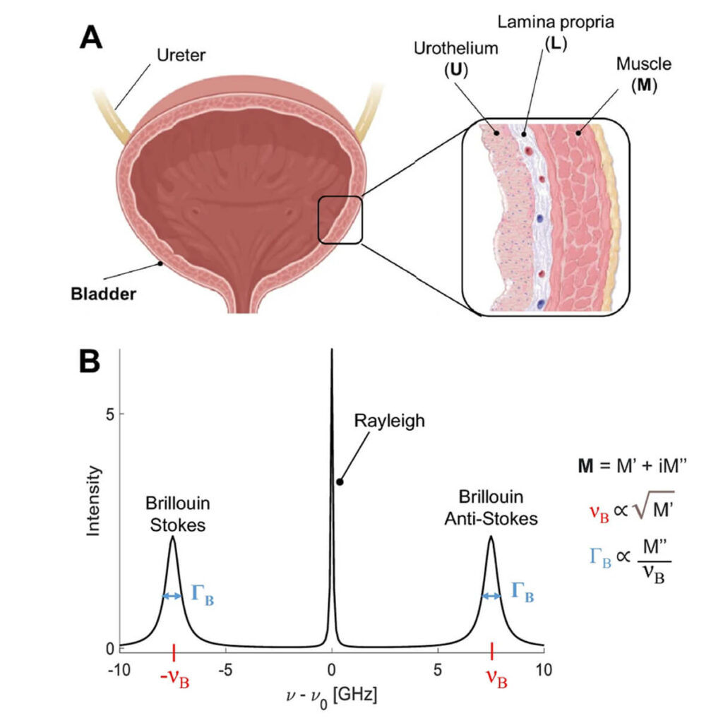
Bladder tissue data
Bladder mechanical properties are crucial for proper organ function and homeostasis, and alterations can indicate disease onset and progression. The research characterizes the tissue elasticity of the murine bladder wall in both healthy and actinic cystitis conditions using Brillouin Microscopy. This technique maps tissue mechanics at the microscale without contact or labeling. In healthy bladders, Brillouin Microscopy distinguished the mechanical properties of different bladder wall layers, with distinct Brillouin shifts for the urothelium, lamina propria, and muscle. In bladders with actinic cystitis, a condition caused by radiation therapy and marked by tissue fibrosis, significant changes in tissue mechanics were observed. Specifically, fibrotic bladders showed lower Brillouin shifts in the lamina propria and decreased mechanical heterogeneity over time, corresponding with fibrosis severity.
Brillouin Microscopy has significant potential as a complementary diagnostic tool alongside histopathological analysis to detect complex mechanical changes in tissues. By providing detailed mechanical mapping, this method could improve understanding of disease progression and tissue remodeling in pathological conditions. The study also compares Brillouin Microscopy with atomic force microscopy (AFM), demonstrating a correlation between the longitudinal modulus measured by Brillouin shifts and the Young’s modulus obtained through AFM, despite differences in measurement principles and frequency ranges. These findings emphasize Brillouin Microscopy’s utility in biomedical imaging and its potential clinical applications for early diagnosis and prognosis of bladder-related conditions.
Source and Copyright:
Martinez-Vidal, Laura, et al. “Progressive alteration of murine bladder elasticity in actinic cystitis detected by Brillouin Microscopy.” Scientific Reports 14.1 (2024): 484.
https://doi.org/10.1038/s41598-023-51006-2
https://creativecommons.org/licenses/by/4.0/


