What is Brillouin Microscopy?
Brillouin Microscopy is a non-invasive, label-free technique that measures the mechanical properties of samples without damaging them. Brillouin Microscopy, in particular, is suitable for biological samples such as cells, biomaterials, tissues, organoids and organisms.
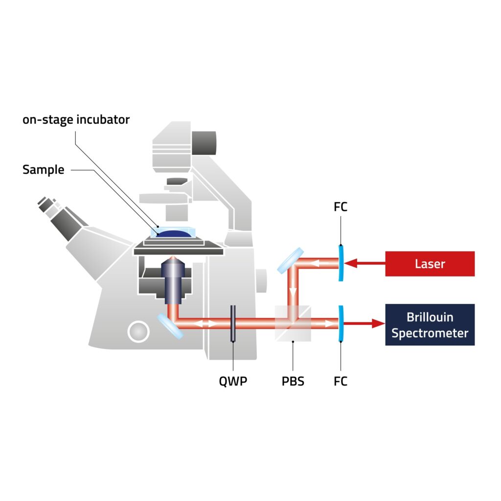
It provides additional contrast channels adding to common microscopy techniques and integrates with research light microscopes. Our Discoverer™ Brillouin microscope adds these functions to existing microscopes enabling correlative studies.
Origin and pronunciation of the term
The method is named after the French physicist Léon Brillouin. Therefore, the right pronunciation is somewhat of a challenge for non-French natives. Here is roughly how it sounds – in phonetic script its [bʁilwɛ̃]:
[bʁ] – Start with a “b” sound, like in “bat.” Immediately after, you’ll make a guttural “r” sound, which is a bit like gargling or the throaty sound you hear in some French words.
[il] – This is pronounced like “eel” in English. It’s a sharp, clear sound.
[wɛ̃] – This final part is a bit tricky. It starts with a “w” sound, like in “we.” The rest of the syllable is similar to the “an” in “can,” but you need to nasalize it. To do this, try to direct the airflow through your nose as you make the sound, giving it a resonant, humming quality.

How does it work? An introduction.
Brillouin Microscopy is an imaging technique that allows scientists to measure the stiffness of cells and tissues without damaging them. It uses light to assess how stiff or soft a material is by observing how light waves are scattered when they pass through it. Specifically, this technique measures changes in the frequency of light (known as the Brillouin frequency shift) caused by interactions with the material, providing information about its mechanical properties. The non-invasive nature of this method makes it ideal for studying delicate biological samples and provides insights into cell behavior and health.
Prevedel, Robert, et al. “Brillouin Microscopy-a revolutionary tool for mechanobiology?.” arXiv preprint arXiv:1901.02006 (2019).
https://creativecommons.org/licenses/by/4.0/
https://arxiv.org/abs/1901.02006
Brillouin Microscopy in more detail
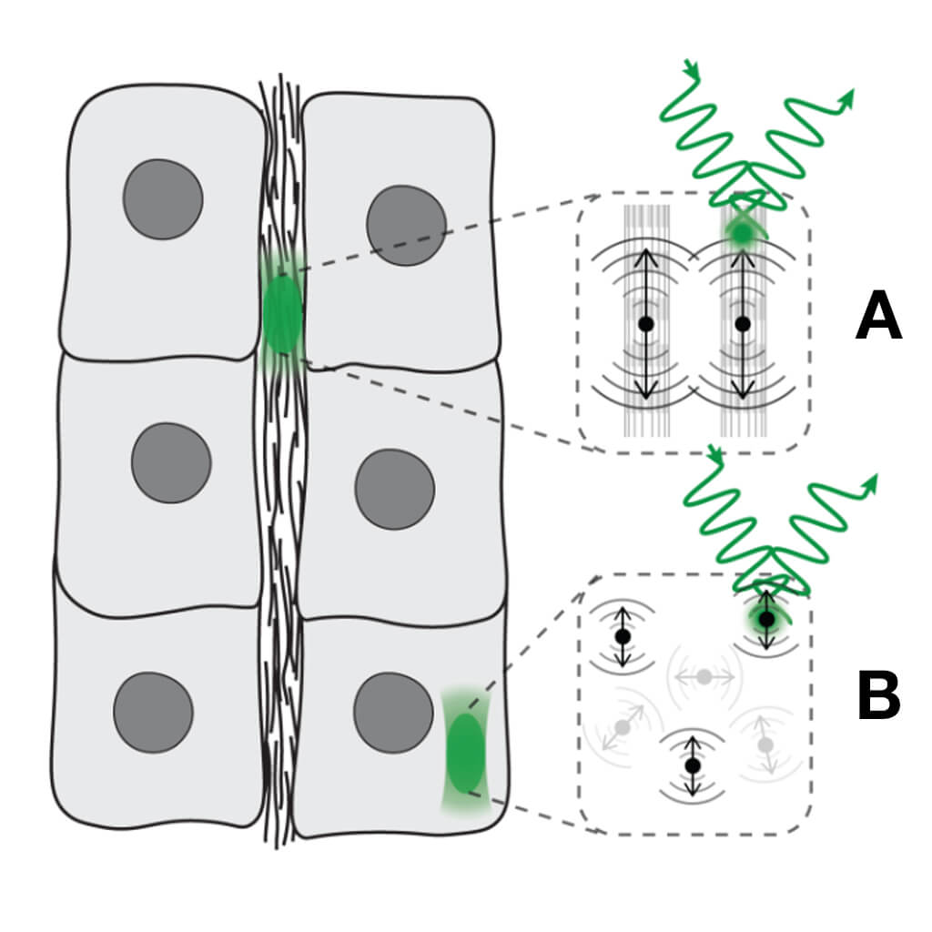
Brillouin Microscopy is based on the principle of Brillouin scattering, where monochromatic light interacts with phonons—quantized sound waves—within a material. When these phonons scatter the incident light, they cause a shift in its frequency, known as the Brillouin shift. This frequency shift is directly related to the acoustic properties of the material, specifically the sound velocity, which in turn can be used to calculate the material’s bulk modulus, a fundamental measure of stiffness.
Brillouin Microscopy is an imaging technique that allows scientists to measure the stiffness of cells and tissues without damaging them. It uses light to assess how stiff or soft a material is by observing how light waves are scattered when they pass through it. Specifically, this technique measures changes in the frequency of light (known as the Brillouin frequency shift) caused by interactions with the material, providing information about its mechanical properties. The non-invasive nature of this method makes it ideal for studying delicate biological samples and provides insights into cell behavior and health.
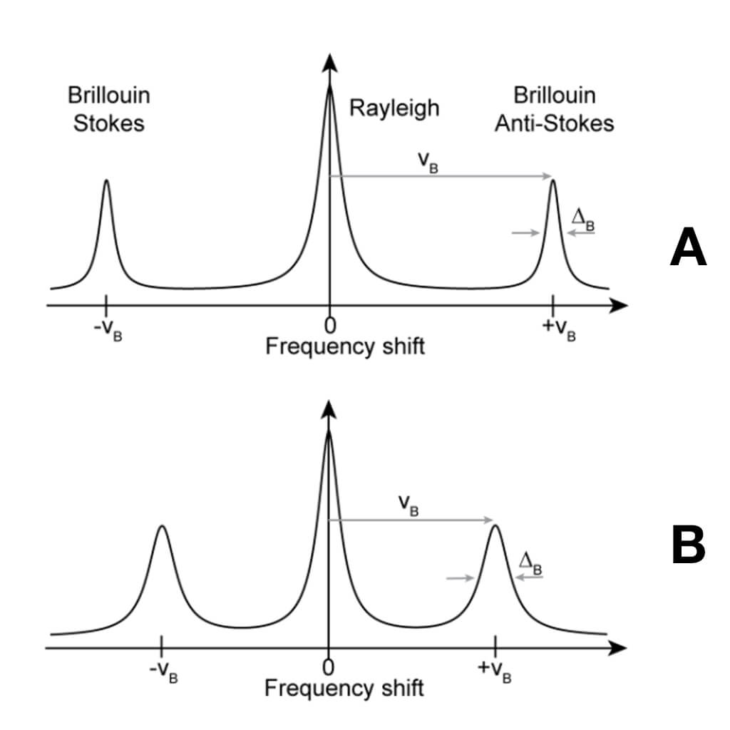
The technique illuminates the sample with a laser, and the light that scatters back contains information about the mechanical environment of the sample due to its interaction with the phonons. By analyzing this scattered light, particularly the frequency and intensity of the Brillouin shift, researchers can deduce not only the stiffness but also other elastic properties of the sample. Since the sound velocity in a material is directly proportional to the square root of the bulk modulus divided by the density, Brillouin Microscopy allows scientists to access these mechanical parameters with high precision.
In practice, Brillouin Microscopy provides a unique advantage in fields like mechanobiology and medical diagnostics, where understanding the mechanical properties at microscale resolutions can be critical. The non-invasive and detailed mechanical insights offered by Brillouin Microscopy complement the chemical and structural information obtained from other microscopy techniques, such as Raman spectroscopy, making it an invaluable tool for comprehensive material characterization. This synergy is particularly useful in studies where both biochemical and biomechanical properties influence biological functions and disease states.
Prevedel, Robert, et al. “Brillouin Microscopy-a revolutionary tool for mechanobiology?.” arXiv preprint arXiv:1901.02006 (2019).
https://creativecommons.org/licenses/by/4.0/
https://arxiv.org/abs/1901.02006
Brillouin Microscopy: 100 years of history
Originating from the work of French physicist Léon Brillouin in the mid-20th century, Brillouin Microscopy emerged as a technique rooted in the principles of inelastic light scattering. In its early applications, particularly in material science, Brillouin Microscopy contributed significantly to understanding the mechanical properties of materials at the microscopic scale. Its roots in solid-state physics laid the groundwork for exploring the mechanical dynamics of materials, ranging from polymers to crystalline matter or nanocomposites.
A notable turning point occurred in the 1990s when researchers recognized Brillouin Microscopy’s potential in mechanobiology, creating a significant expansion of its scientific relevance. The subsequent decades saw an evolution that extended beyond its initial focus on materials. Scientists have harnessed its capabilities to delve into the biomechanics of cells and tissues, studying stiffness variations on microscopic scales. In more recent times, Brillouin Microscopy has found noteworthy applications in plant physiology.
Throughout the 21st century, ongoing technological refinements pushed Brillouin Microscopy into a prominent position within the scientific community. Enhanced resolution and increased adaptability strengthened its status as a practical and non-invasive method for approaching the mechanical intricacies of living cells, tissues, materials, and even the physiology of plants. In its evolution, Brillouin Microscopy became integral to a spectrum of disciplines, bridging the realms of physics and biology, and leaving a mark in both material science and life science research.
CellSense brings Brillouin Microscopy to new users and application fields by creating a user-friendly solution that can be easily operated without requiring deep expertise in physics or spectroscopy. Our mission is to simplify the technology, overcoming technical complexity to make it accessible to a broader audience.
A Lively and Growing Community
Brillouin microscopy has moved well beyond the physics lab. Today, a broad and fast-growing user community applies it across the life sciences—from cell and tissue mechanics to developmental biology and plant research. What began as a specialized optical method is now becoming a practical tool for studying living systems, providing direct access to mechanical information without labels or contact.
The BioBrillouin Network connects scientists from many disciplines who share data, methods, and experience. Regular meetings, workshops, and collaborations keep the field moving forward and make it easy for newcomers to get involved. It’s a dynamic, open community that drives innovation and keeps Brillouin microscopy close to real biological questions.